Clinicians need to act quickly to administer effective treatment when a patient has an acute ischemic stroke — the most common type — which occurs when a clot blocks blood flow through an artery in the brain. Often, this involves conducting a brain scan to map the damage caused by the stroke — either using magnetic resonance imaging (MRI) or computed tomography (CT).
But these scans expose patients to chemical contrast agents, which can be problematic. Some contain high doses of radiation, while others can be dangerous for patients with kidney or vascular problems.
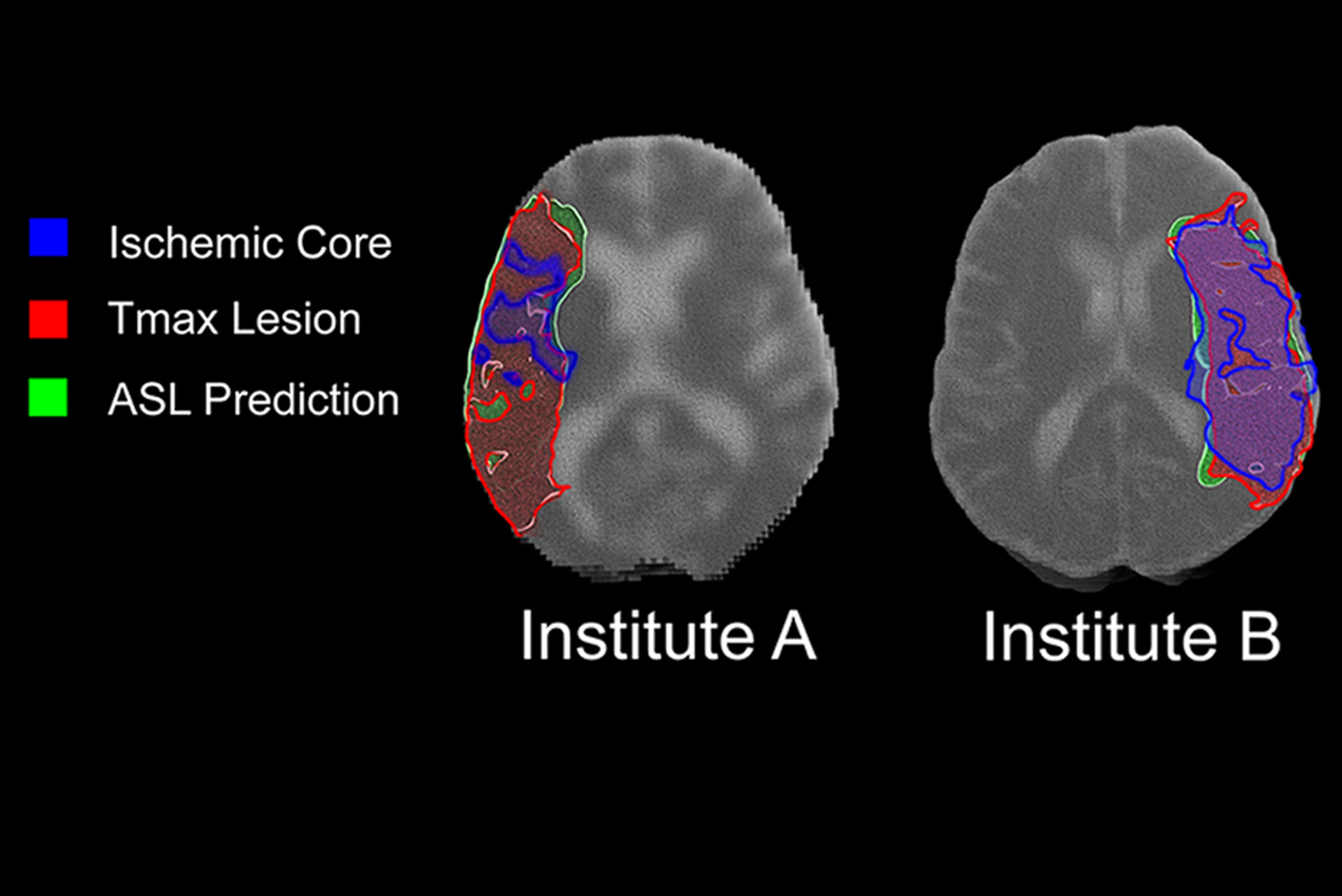
Researchers at the USC Mark and Mary Stevens Neuroimaging and Informatics Institute are working on an alternative that allows clinicians to assess stroke damage without injecting patients with contrast agents. In a new publication, published in the journal Stroke in December, a team lead by researchers at the institute built and tested an artificial intelligence (AI) algorithm that can extract data about stroke damage from a much safer type of brain scan known as pseudo-continuous arterial spin labeling (pCASL) MRI.
“This is the first systematic study that applies a ‘deep learning’ algorithm and non-contrast perfusion MRI to identify brain tissue damaged by a stroke across platforms and institutions,” said Danny JJ Wang, PhD, professor of neurology at the USC Stevens Neuroimaging and Informatics Institute and last author of the study. “Our model is a promising approach to assist clinical decision-making for stroke patients — and it’s completely non-invasive.”
Wang and his team at the institute — including PhD candidates Kai Wang, Qinyang Shou and Samantha Ma, and Hosung Kim, PhD — collaborated with scientists at the University of California, Los Angeles (UCLA) and Stanford University to conduct the study. To train their algorithm, the researchers used 167 sets of images, collected on 1.5Tesla and 3.0Tesla Siemens MR systems from 137 patients with acute ischemic stroke at UCLA. The trained model was independently validated on 12 sets of images collected on 1.5Tesla and 3.0Tesla GE MR systems at Stanford.
Their results are extremely promising. The pCASL deep learning model had an accuracy of 92% in assessing damaged brain tissue in tests of two separate groups of stroke patients.
“A highlight of this finding is that we’ve shown this method is effective across institutions, MR platforms and cohorts,” Wang said.
Next, Wang plans to conduct a larger-scale study to evaluate the algorithm at additional institutions and to extend the treatment window for acute ischemic stroke beyond 24 hours after the onset of symptoms.
“At the INI, we’re investing significant resources in developing AI tools to aid neurologists in predicting, diagnosing and treating various types of neurological dysfunction,” said Arthur W. Toga, PhD, director of the institute. “Dr. Wang’s recent breakthrough is an important step toward this broader goal.”
— Zara Greenbaum