A team of scientists at the Keck School of Medicine of USC is looking to some deep sea dwellers to create a better way to develop cancer-fighting therapies. Harnessing the power of the enzymes that give these marine animals the ability to glow, the team created a test that makes it easy for researchers to see whether a therapy is having its intended effect — killing cancer cells. The results of their study were published in Scientific Reports on Jan. 9.
“One of the most promising areas in cancer research is immunotherapy, including chimeric antigen receptor-T (CAR-T) cells. It is also one of the most difficult because the methods for testing immunotherapies are not ideal,” said corresponding author Preet M. Chaudhary, MD, PhD, professor of medicine at the Keck School, chief of the Jane Anne Nohl Division of Hematology and Center for the Study of Blood Diseases, and director for bone marrow transplant at USC Norris Comprehensive Cancer Center. “Radioactive chromium release assay is the gold standard for testing whether an immunotherapy kills cancer cells. This method is expensive, complicated and requires special disposal practices. Other available methods also suffer from limitations and don’t allow scientists to rapidly screen immunotherapeutic agents to find the best candidates.”
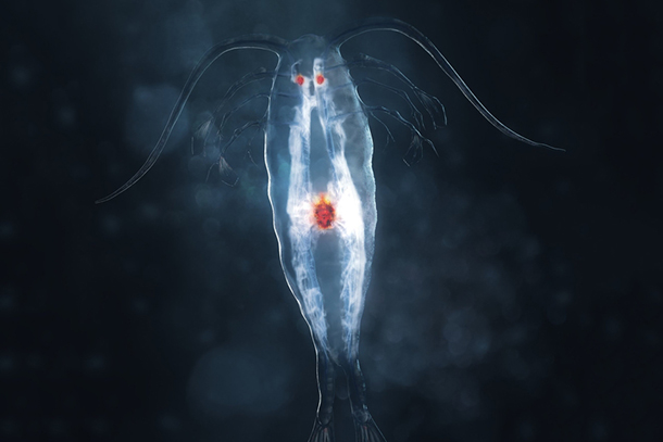
The team set out to develop a simple, precise and inexpensive test based on marine animal luciferases, the enzymes responsible for bioluminescence. A group of small crustaceans and deep sea shrimp were selected for their bright bioluminescence, and their luciferases became the basis of the test, called the Matador assay. Engineered to get trapped inside cells, the luciferases leak out of cells when they die, causing a visible glow. The level of luminescence can then be measured with a luminometer.
To test the Matador assay’s effectiveness at measuring cell death, several different types of cancer cells, including chronic myelogenous leukemia, acute myelogenous leukemia, Burkitt’s lymphoma and solid tumors, were treated with a variety of immunotherapies, including CAR-T cells, bispecific T-cell engagers and monoclonal antibodies. Results showed that the assay was so sensitive that it could detect the death of a single cell, a level of sensitivity far exceeding existing assays. Chaudhary’s lab has since developed more than 75 cancer cell lines expressing the marine luciferases and used them successfully in the Matador assay to develop next-generation CAR-T cells.
The assay is fast, inexpensive and can be performed in a 384-well plate format, saving time and reagents.
“In our hands, the Matador assay can detect cell death in as little as 30 minutes, which can ultimately translate to more expedient treatments for patients getting cellular immunotherapies such as CAR-T cells,” Chaudhary said.
The Matador assay has many potential applications in biomedical research and cellular therapy manufacturing, he explained.
“It could potentially play a role in screening other types of anticancer agents or even measuring environmental toxins.”
Hittu Matta, PhD, assistant professor of research medicine, Ramakrishnan Gopalakrishnan, PhD, research associate, Sunju Choi, PhD, research associate, Rekha Prakash, MS student, Ruben Prins, research technician, Venkatesh Natarajan, assistant professor of research medicine and Sonjie Gong, research technician, among others, also contributed to this study.
— Erica Rheinschild