The USC Mark and Mary Stevens Neuroimaging and Informatics Institute (INI), part of the Keck School of Medicine of USC, has built and launched a powerful new smartphone application, Schol-AR, that uses augmented reality (AR) to revolutionize how research and educational outputs are shared. In the most extensive effort to apply AR technology to scientific visualization yet, Schol-AR allows scientists to embed 3D models, videos, and other data within posters, publications and presentation materials to improve engagement and the explanatory power of their work.
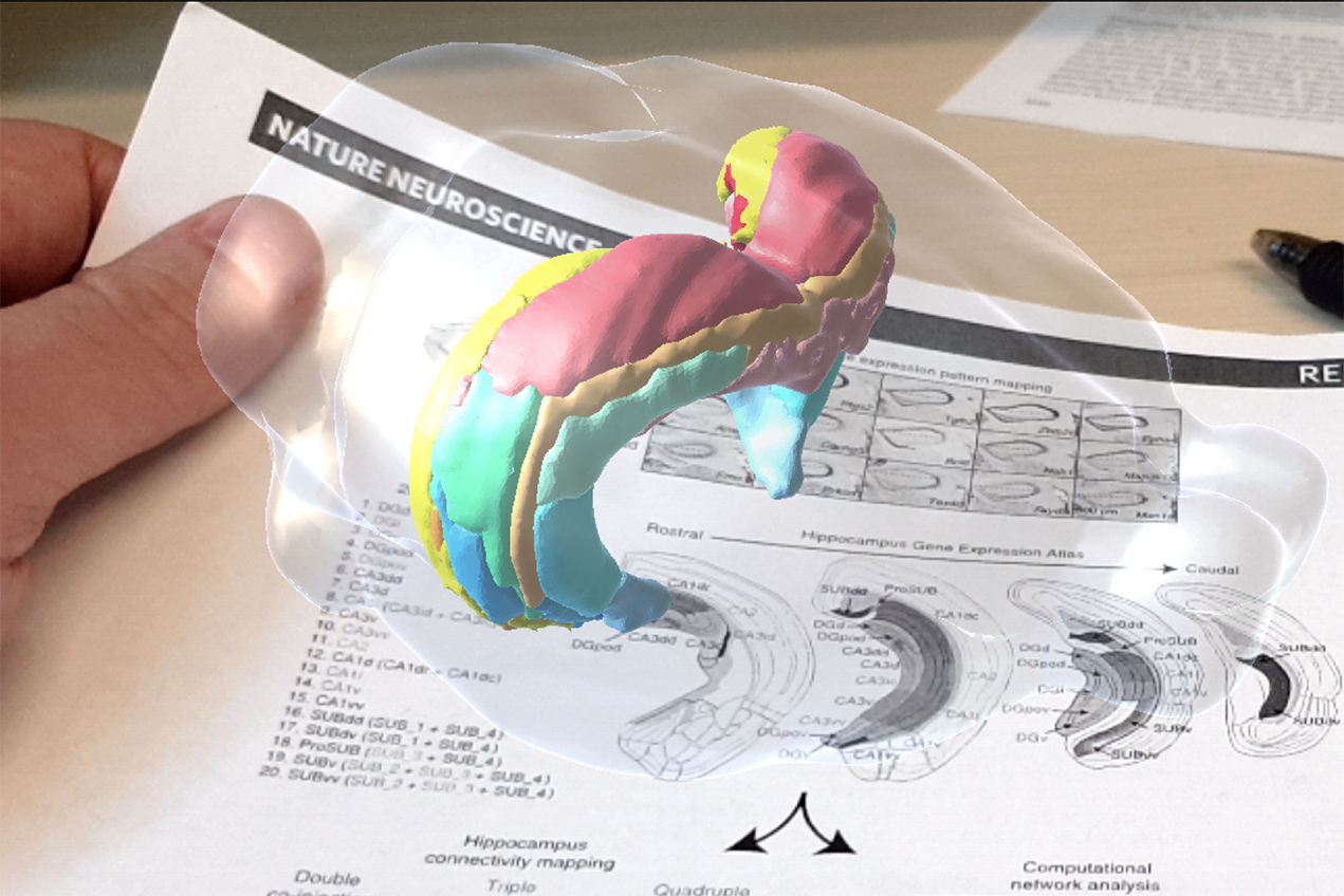
The new application, created by Tyler Ard, PhD, assistant professor of research neurology at the Keck School, was built using software similar to that which helped create video games such as Pokemon Go. Users who have downloaded the application simply point their smartphone cameras at a supported image to pull up hidden interactive content. With the potential to significantly transform how science is communicated, Schol-AR soon will be available for use by researchers across USC and beyond.
“Our goal is to facilitate the exchange of ideas and information in a way that’s less prone to misinterpretation, and more conducive to conveying the deep comprehension that underlies the scientific process,” Ard said.
Schol-AR debuted on the cover of the journal NeuroImage’s October issue, which features a publication by Danny JJ Wang, PhD, professor of neurology and radiology at the Keck School and director of imaging technology innovation at the INI. The 2D cover image, based on high resolution data collected by the INI’s ultra-high-field 7Telsa magnetic resonance imaging scanner, depicts very small blood vessels in the brain, known as lenticulostriate arteries (LSAs). Wang and his team have developed and tested a new non-invasive method for precisely visualizing LSAs, which can help in the study and treatment of cerebral small vessel disease.
However, such 2D images provide little to no information about how LSA size, shape and positioning relates to the brain structures they supply. By layering a 3D model on top, which users can manually enlarge, rotate and explore, a much more realistic picture of the underlying biology emerges.
“The 3D model provides more insight into where these arteries are situated within the context of important structures in the brain,” said Samantha Ma, a graduate student researcher at INI, first author on the study and a member of Wang’s team. “Interacting with the augmented version allows readers to understand how the shape of these vessels could change with age, disease or both.”
Schol-AR will be featured at the annual meeting of the Society for Neuroscience in Chicago, Oct. 19-23. Soon after, the INI will release Schol-AR Creator, a companion application that allows researchers everywhere to upload and embed interactive figures within their own educational materials.
“From its inception, we’ve opted to democratize this technology to aid the efforts of researchers across scientific disciplines,” said Arthur W. Toga, PhD, Provost Professor of Ophthalmology, Neurology, Psychiatry and the Behavioral Sciences, Radiology and Engineering, the Ghada Irani Chair in Neuroscience at the Keck School, and director of the INI. “We envision that this groundbreaking new way of visualizing scientific data will change the way findings are communicated well beyond the field of neuroscience.”
To download Schol-AR, go to www.ini.usc.edu/ScholAR/download.html.
— Zara Greenbaum