For patients with tumors, strokes, cerebrovascular disease and other conditions, computed tomography (CT) scans can be lifesaving. But there’s a growing concern that the radiation involved is unnecessarily harmful to patients. It’s estimated that for every 2,000 CT scans, one future cancer case will occur — which translates to 50,000 cancer cases from the 100 million scans performed in the U.S. each year. A team based at the Keck School of Medicine of USC is working to change that.
“Our technology reduces the radiation dose of CT perfusion by about 75% without compromising imaging speed or quality,” said Danny JJ Wang, PhD, director of imaging technology innovation at the USC Mark and Mary Stevens Neuroimaging and Informatics Institute (INI). CT perfusion is a widely used CT exam that measures blood flow to the brain using a high dose of radiation.
In August, Wang and his team received a two-year, $1.6 million grant from the National Institutes of Health’s Small Business Innovation Research program for the second phase of their project. The program supports the translation of technologies from the lab to commercial use through small business innovation and commercialization.
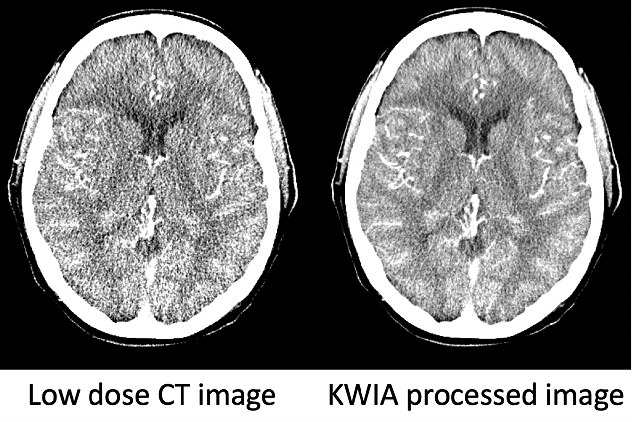
Compared to existing low-dose CT technologies, the team’s new technique, known as K-space Weighted Image Average (KWIA), has the advantages of fast computation speed and excellent retention of the texture and resolution of CT images, as shown in the image above.
“KWIA dramatically improves the quality and signal-to-noise ratio of low-dose CT images, with minimal additional computation time,” said Vinay Duddalwar, MD, professor of clinical radiology at the Keck School. “The technology may impact the safety and clinical practice of CT perfusion.”
Wang and his colleagues published their first study of KWIA in IEEE Transactions on Medical Imaging, a journal of the Institute of Electrical and Electronics Engineers, in July. The study tested the KWIA method and found that the image quality, spatial and temporal resolution and accuracy of low-dose CT perfusion scans were comparable to traditional CT methods, while the radiation dose was reduced by up to 75%.
The INI is collaborating with USC’s radiology department, including Duddalwar, Chia-Shang (Jason) Liu, MD, and Jeffry Alger, PhD, at Hura Imaging, Inc. Duddalwar and Liu are evaluating the clinical utility of this technology, the INI provides the facilities and computational resources through its Laboratory of Neuro Imaging (LONI) and Hura Imaging is handling commercialization of the technology.
“Dr. Wang is leading the institute’s efforts to use our computational resources and expertise in clinical imaging to make CT scans safer,” said Arthur W. Toga, PhD, Provost Professor of Ophthalmology, Neurology, Psychiatry and the Behavioral Sciences, Radiology and Engineering, the Ghada Irani Chair in Neuroscience at the Keck School, and director of the INI.
In addition to conducting additional research on KWIA and other techniques, the team is applying for clearance from the U.S. Food and Drug Administration.
“Our long-term goal is to commercialize this tool so that it can be applied as widely as possible to clinical practice,” Wang said.
— Zara Greenbaum