This colorful series of mouse skulls reveals stem cells, labeled with the protein Gli1, in the sutures between the calvaria bones in the upper part of the cranium.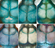
Among other things, these stem cells support the postnatal turnover and injury repair of the calvaria bones.Hu Zhao, DDS, PhD, a research associate in the lab of Yang Chai, DDS, PhD, in the Center for Craniofacial Molecular Biology at the Ostrow School of Dentistry of USC, produced the image, which won the December 2013 USC Stem Cell Image of the Month Contest. The contest invites USC stem cell researchers to submit high-resolution images or artistic renditions that showcase the scientific excellence and creativity of the university’s research enterprise. Submit images, one-sentence informative captions, names and laboratory affiliations to Seth Ruffins, PhD, at ruffins@usc.edu by the last day of each month to enter.