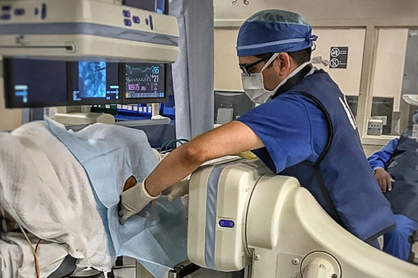
On July 1, Vishal Patel, MD, PhD, joined the Mark and Mary Stevens Neuroimaging and Informatics Institute (INI) at the Keck School of Medicine of USC as an assistant professor of radiology. Patel, who earned his doctoral degree in biomedical engineering, completed a residency in diagnostic radiology at the University of California, Los Angeles and a fellowship in neuroradiology at USC.
Patel’s research interests include MRI, diffusion imaging, tractography and machine learning. At the INI, he plans to apply his knowledge of computational MRI and artificial intelligence (AI) to improve diagnostic techniques for neuroradiologists.
“The goal is to establish USC, and in particular the INI, as a site where AI methods and techniques are developed, validated and made available for use to the greater neuroimaging community, including both researchers and clinicians,” Patel said.
AI holds promise for a number of clinical challenges. For instance, an MRI scanner may register images of very small brain lesions that the naked eye cannot detect, but researchers like Patel can build software that identifies subtle abnormalities present in the underlying imaging data. Ultimately, this technology can be used to detect tumors and classify them by cell-type and pathological grade, providing a precise and non-invasive way to diagnose brain cancer.
Patel’s research at USC will build on his previous work applying machine learning concepts to tumor identification and other clinical challenges. But now, he has a powerful new tool: the INI’s ultra-high field 7Tesla MRI scanner, which received FDA approval for clinical scanning last fall.
“The INI is proud to welcome Vishal to our team,” said Arthur W. Toga, PhD, provost professor at USC and director of the INI and the Laboratory of Neuro Imaging (LONI). “Applying his expertise in neuroradiology to our high-end clinical imaging system will undoubtedly lead to meaningful new discoveries.”
Toga and Patel have collaborated in the past and look forward to resuming their research together on a new clinical imaging study of patients with multiple sclerosis (MS).
“LONI was a phenomenal place during my graduate training and it continues to become more and more impressive, especially with the addition of clinical high-field imaging capabilities,” Patel said. “I’m excited to come on board and get to work in what I know to be an excellent environment for research.”
— Zara Greenbaum


