“All these things we do, it’s X-ray beam interaction with matter in order to visualize structures of different materials,” said Tea Jashashvili, MD, PhD, assistant professor of research radiology at the Keck School of Medicine of USC, as she described the work that goes on at the nano- and micro-CT lab of the USC Molecular Imaging Center (MIC) on the Health Sciences Campus. At its inception, the facility was tasked with handling small animal imaging, but today the MIC has grown into one of the nation’s premier imaging resources, in large part due to major equipment funds from the National Institutes of Health and USC.
This continued expansion has benefited Keck School researchers as well as those outside of USC. The most recently added instrument capabilities include ex-vivo micro- and nano-CT imaging, and have proven invaluable to the work of these researchers. “Today, basic, pre-clinical or biological research is not only supplemented by nano- and micro-CT data, but increasingly often it provides the actual raw data serving as evidence to advance science in these respective fields,” Jashashvili said while displaying several images of nanostructures of different materials acquired using 3D nano-CT.
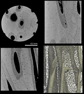
Microstructural details of cortical bones are visualized using the Nanotom-GE system, scanned at 0.70 µm pixel size resolution. X-ray images are produced as transverse (upper left), sagittal (lower left), and coronal (upper right) sections of a bone sample with a 3-D rendering of its Haversian canal and lacunae network (bottom right). (Illustration/Courtesy Tea Jashashvili)
At the MIC, faculty and staff work toward developing advanced imaging methods to service the USC research community and the larger scientific community in general. Along these lines, the MIC’s improved staining method, using phosphate-buffed saline and phosphotungstic acid (PBS/PTA), binds high-atomic elements to soft tissue structures, taking advantage of the osmotic differences that draw higher-density material in and allow for enhanced X-ray images of low-density soft tissue. Because of a staining protocol devised at the MIC, low-density soft tissue is identifiable when using high resolution computed tomography (micro-CT) technology.
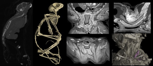
For example, “a startlingly detailed series of images of a tarsier provided by the center offers proof of this method’s ability to map soft tissue that would not otherwise be dense enough to be visible by X-ray,” said Tautis Skorka, MPH, systems analyst at MIC, when describing a recent scan of a tarsier, which is a small primate located in Southeast Asia. This methodological improvement, along with others, offers the possibility to non-destructively study structures of interest and, importantly, return specimens to museum collections or research labs from which they were borrowed so that they subsequently can be used for other applications.
Through combining an array of different imaging instruments (MRI, microPET, Ultrasound/Photoacoustics, and optical imaging) with innovative methods conceived to improve 3-D imaging, MIC, as a core facility, seeks to serve the diverse research needs of the entire USC community. For example, “Researchers at the Natural History Museum rely on MIC equipment and support for important research that entails CT scanning a diversity of fossils, from large dinosaurs to small feathers embedded in ancient amber. We are thrilled to have this collaboration with the MIC,” said Luis Chiappe, PhD, adjunct professor of earth sciences and biological sciences at USC, as well as senior vice president of research and collections and Gretchen Augustyn Director of the Dinosaur Institute at the Los Angeles County Natural History Museum.
The MIC is available to users from USC or other institutions, and always welcomes new projects.
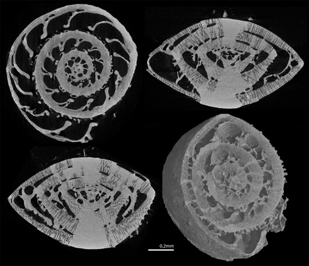
Microstructural details of marine shell walls are visualized using the Nano3DX XRM-Rigaku system, scanned at 0.27 µm pixel size resolution. X-ray images are produced as transverse (upper left), sagittal, (lower left), and coronal (upper right) sections of the obliquely sectioned 3-D model (bottom right). (Illustration/Courtesy Tea Jashashvili)