Neuroscience researchers have long used MRI to study the basic structure of the brain in health and disease, but these scanners typically cannot provide insights at the cellular level. In an MRI image, each voxel — the 3D equivalent of a pixel — represents tens of thousands of brain cells.
But a breakthrough study led by the USC Mark and Mary Stevens Neuroimaging and Informatics Institute (INI) at the Keck School of Medicine of USC has shown that it’s possible to use high-resolution MR imaging and a novel cell labeling technique to visualize less than 100 cells. The results ultimately will allow clinicians to assess the effectiveness of cell-based therapies used to treat various cancers.
“The application of this new method is very broad,” said Danny JJ Wang, PhD, professor of neurology and radiology at the Keck School, adding that it can be used to monitor both immune and stem-cell therapies. “We used standard clinical equipment and FDA-approved drugs in the hopes that the results can translate quickly from bench to bedside.”
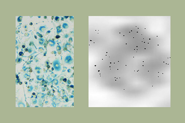
In the study, researchers tested a new method of labeling cells, which they correctly predicted would render cells easier to visualize in an MRI scanner. They labeled macrophages, a type of white blood cell involved in the immune response, with three FDA-approved compounds — heparin, protamine and ferumoxytol. The team then used the INI’s cutting-edge MRI scanners, including the new ultra-high magnetic field 7T Terra, to capture images of the macrophages. In some trials, they were able to detect as few as 62 cells labeled with the three compounds.
“One of our missions at INI is to work alongside clinicians to solve intractable problems facing the modern medical community,” said Arthur W. Toga, PhD, Provost Professor of Ophthalmology, Neurology, Psychiatry and the Behavioral Sciences, Radiology and Engineering, Ghada Irani Chair in Neuroscience and director of the institute. “Dr. Wang’s team is committed to advancing that mission by expanding the reach of our powerful imaging equipment.”
The researchers involved in the study include Wang’s team at INI, along with Wange Lu, PhD, associate professor of stem cell biology and regenerative medicine at the Keck School, and Yelong Shen, MD, from the Shandong Medical Imaging Research Institute at Shandong University in China. The paper was published July 3 in the International Journal of Nanomedicine.
— Zara Abrams