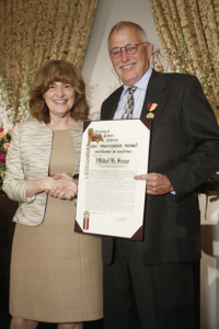
Elizabeth Graddy presents Mikel Snow with the USC Associates Award for Excellence in Teaching during the Academic Honors Convocation, held April 17 at Town and Gown on the University Park Campus. (Photo/Steve Cohn)
Mikel Snow, PhD, is professor of cell and neurobiology (educational scholar), and holds the Lt. Col. Earle and Patricia M. Smith Professorship in Neurogenetic Research at the Keck School of Medicine at USC.
On April 17, Snow was presented with the 2017 Associates Award for Excellence in Teaching at the university’s 2017 Academic Honors Convocation.
The Associates Award for Excellence in Teaching is the highest honor the university’s faculty members present. It recognizes the career achievements of outstanding faculty who have a proven track record as exceptional teachers, have a positive, inspiring, and long-lasting effect on students and their learning, and extraordinary, unique, or pioneering contributions to excellence in teaching.
Snow recently sat down with HSC News to discuss his path to teaching and his work in the field of cell and neurobiology.
Question: What drew you to teaching instead of simply conducting research?
Answer: When I started my career, I had an active research program on the origin of regenerating muscle fibers resulting from injury or disease. Following a sabbatical leave at the University of Washington to learn molecular biology and tissue culture of human muscle cells, I was asked in 1990 to be the assistant dean for student affairs at USC. Since accepting this position and continuing to teach and direct one of the largest courses (anatomy) at the medical school, my time for research was severely curtailed. Thus, this marked the beginning of my career change to focus more on teaching and administration.
Q: How has medical education changed in the years that you have been at the Keck School?
A: The biggest change has been in the basic sciences (first two years), with a change in emphasis from details (minutia) in favor of an increase in more clinically relevant basic science, coupled with an improvement in integration across the basic sciences.
Q: You’ve been described as “the most decorated basic science educator in the history of the Keck School of Medicine.” What does it take to be a great teacher?
A: A great deal of hard work to perfect and upgrade lecture presentations, taking full advantage of the latest technologies available. In addition, the best teachers really care about the students and how they best learn, since “one size does not fit all.” In my discipline, this requires considerable time working with students one-on-one outside the classroom to show them a more efficient and effective way to learn the material. Equally important is being very knowledgeable about the subject; in anatomy, being a skilled dissector is as important, or more important, than being an excellent lecturer. To this end and for several years following my training, I continued to hone my skills by dissecting a body before each lab. Finally, an important component is having an approach, or personality, that inspires student to learn and enjoy the subject, an approach that is not intimidating. Medical students want to be great doctors, so encouraging and supporting them is much more effective than scaring them.
Q: What are some of the advances in cell and neurobiology that get you and your students excited about the field?
A: Speaking from my role as a teacher, I’ve found that human anatomy is often thought of as a static or non-changing field — this is not true. For example, I did some research last year with a pediatric surgeon colleague trying to develop a better way to remove thyroglossal cysts in the neck. Advances in imaging technology have had a huge impact on teaching human anatomy. Many years ago, radiology was not a significant component to anatomy courses. I received no radiology exposure in the anatomy course I took as a graduate student, so I taught myself basic radiology, which is now an integral part of our Keck School anatomy course, and which is obviously very exciting to students. More recently, ultrasound has been added to our course, as yet another way to visualize the body that has clinical relevance.
Q: What do you see coming in the field? What advances do you hope your students may achieve?
A: We are in the digital age, so many companies are developing software for teaching anatomy. For example, I am heading out this afternoon to visit a facility in Irvine to evaluate virtual reality software on viewing the body with holograms. Many of these products are ready for the classroom for allied health training programs. They also have great potential for teaching medical students, but not as a replacement to dissecting a donor, which continues to be a hands-on experience that effectively teaches 3-D relationships, an appreciation for bodily variations, and the layering of tissues surrounding the body parts.
— Leigh Bailey