When Andy McMahon, head of USC Stem Cell, wanted a three-dimensional image of a kidney, he used to ship the organ to Australia. Now he can just send down the hall to the university’s new specialized microscope — built by five undergraduates from the USC Viterbi School of Engineering for ENGR 499 Microscope Design and Construction.
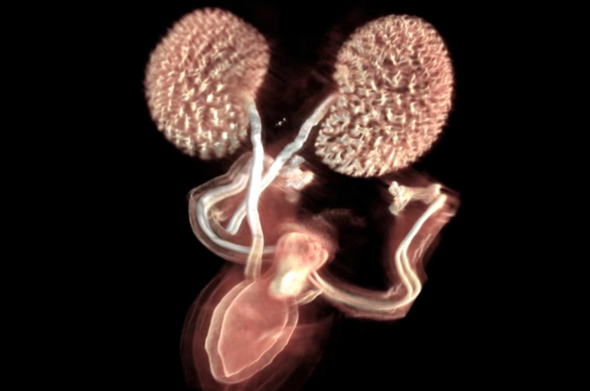
Called an optical projection tomography (OPT) microscope, the instrument produces 3-D images of pea-sized biological samples such as organs and embryos. This provides a valuable tool that enables McMahon and other biologists to study how organs develop and how they maintain and repair themselves.
“To acquire an OPT image, light is either projected through the specimen or the specimen is illuminated for fluorescent imaging. Then a camera captures images at 1,000 different angles all the way around the specimen,” said Seth Ruffins, who taught the course and directs the Microscopy Core Facility at the Eli and Edythe Broad Center for Regenerative Medicine and Stem Cell Research at USC. “And through a mathematical process called a back plane projection you can then construct those images from all these different angles into a three-dimensional shape.”
With funding secured by McMahon and input from USC Viterbi Professor Andrea Armani and Provost Professor Scott E. Fraser, the undergraduates built and assembled the microscope hardware in ENGR 499 during the fall 2014 semester. They continued to troubleshoot the software as volunteers during spring 2015. Chao-Yuan “Joe” Yeh, a graduate student in the lab of Cheng Ming-Chuong at the Keck School of Medicine of USC, further refined the instrument control software.
The microscope began capturing its first state-of-the-art images, including a detailed three-dimensional rendering of kidneys and lungs, in June.
“This is probably only the fourth or fifth microscope of this design built in the United States,” said Ruffins. “This particular design comes from a group in Toronto [that] built it as an open source project.”
Even with those designs in hand, the process of building the OPT microscope presented unexpected challenges.
“It wasn’t a step-by-step blueprint at all, so we had to fill in the gaps and a lot of blanks there,” said Stephanie Fong, a biomedical engineering major with a digital studies minor. “So, in a sense, this was more representative of the real world than a classroom situation. It was really just jumping in and getting hands-on right away with very little prior knowledge.”
Biomedical engineering major Dinesh Seemakurty added: “It shows you that, even with a plan, it’s still very difficult to make something happen. There are bugs, or there’s something that goes wrong. And no one actually has documented how you solve that, so you end up having to figure that part out.”
The students did everything from machining the parts to building the microscope’s computer, plus assembling the components and programming software.
“I have to say it’s a very different course,” said Beibo Zhao, a biomedical engineering major with a statistics minor. “You have to get your hands dirty to do stuff, to learn to make a computer. So that’s something very valuable.”
The hands-on nature of the course taught the students a great deal in a short time. In the process, they discovered how to work as a team to solve a real-world problem on a tight deadline.
“We’re all biomedical engineers, but we all have different strengths,” said Kathleen Roche, a biomedical engineering major with a French minor. “In most of our classes, we don’t really get the chance to build a machine, especially something as complicated as this from the ground up, so it’s been a really cool experience. We learned how to be innovative.”
McMahon will continue to cultivate opportunities for collaboration and interdisciplinary thinking through the university-wide USC Stem Cell initiative, which connects nearly 100 researchers from all disciplines.
“I look forward to continuing to work with students and colleagues at USC Viterbi and across the university,” he said. “The fact that this undergraduate course has produced a working OPT microscope, which will be tremendously useful to stem cell researchers including myself, underscores the synergy that results when we reach across disciplines.”
— Cristy Lytal