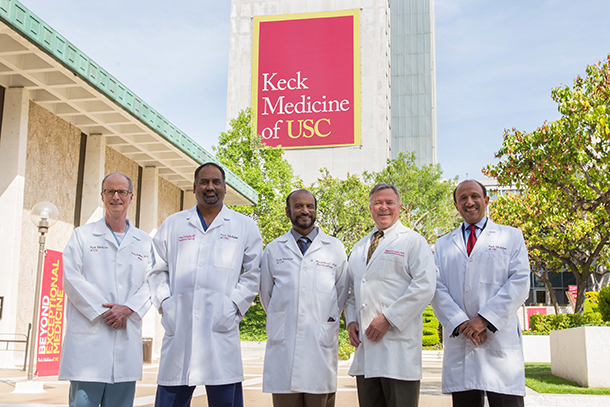
A surgical team at Keck Medicine of USC pushed the boundaries of clinical care by performing the first-ever robotic, minimally invasive surgical removal of a stage IV tumor thrombus — a kidney cancer tumor that extends into the heart.
The nearly 10-hour procedure required painstaking precision from three renowned surgeons, a critical care anesthesiologist and a radiologist. In doing the procedure, the team reduced the patient’s risk of sudden death from the tumor breaking off into the heart and lungs.
Typically, the surgery for a stage IV tumor thrombus, or blood clot, is both traumatic and risky. It requires major open surgery in which the patient’s chest and abdomen are opened completely while the anesthesiologist monitors the patient and the thrombus. The aim is to remove the tumor and thrombus from the inferior vena cava and the heart while ensuring it does not break. Several quarts of blood are needed for transfusion and patients have a 1 in 20 chance of dying during the procedure.

Animated 3-D imaging of the patient’s organs was used to plan the complex procedure. (Illustration/Courtesy of Inderbir Gill)
The use of robotic surgery techniques significantly reduced trauma to the patient and minimized blood loss by more than five-fold. Because the incisions were small, the patient’s hospital stay was just six days, as opposed to the typical two to three weeks after open surgery. Overall recovery time was also reduced significantly.
Such multidisciplinary collaboration lays the groundwork for using advanced technology to build higher standards of patient care, even in the most complex cases.
“This exciting feat promises to redefine the boundaries of what is surgically possible through skill, collaboration and technology,” said Inderbir Gill, MD, Distinguished Professor and chair of urology, founding executive director of the USC Institute of Urology and associate dean for clinical innovation at the Keck School of Medicine of USC. Gill led the multidisciplinary team that performed the surgery. “Our hope is that we can now propel the field at large to turn such futuristic robotic surgery into our present standard of care.”
Pinpoint precision
Before the surgery, Vinay Duddalwar, MD, associate professor of clinical radiology, created three-dimensional animated maps of the patient’s chest and abdomen so that surgeons could pre-plan their entire surgical strategy with millimeter precision. The procedure began with Namir Katkhouda, MD, PhD, professor of surgery, performing a surgical maneuver to control blood flow to the patient’s liver. Next, Gill used the latest-generation Xi da Vinci surgical robot to completely dissect the tumor-bearing kidney through small keyhole incisions in the patient’s abdomen to control various blood vessels, which allowed him access around and into the inferior vena cava where the cancer had spread. Then Mark Cunningham, MD, associate professor of clinical surgery, put the patient on a heart-lung bypass machine to create a bloodless environment for tumor removal. He opened the patient’s heart using a minimally invasive incision through the rib cage.
Cunningham and Gill then worked quickly and simultaneously from the chest and abdomen to remove the tumor thrombus from the heart and inferior vena cava, respectively, with Cunningham working from the chest downward and Gill working from the abdomen upward. All the while, Duraiyah Thangathurai, MD, professor of clinical anesthesiology and chief of critical care medicine, monitored the patient’s organ function, keeping a close eye on the patient’s heart using an esophageal echo probe. If a portion of the tumor were to break off into the heart or lungs, the patient would die instantly.
“We are proud of our ability to coordinate such complex efforts between the cardiac and urologic surgical teams with skill and dexterity,” Cunningham said. “This was the driver of our success and exactly the standard we strive for across the institution.”
According to the American Cancer Society, there are approximately 64,000 new cases of kidney cancer diagnosed each year. Only a small fraction of those patients (about 10 percent) have cancer that spreads through the blood vessels without metastasizing to other organs. In a small portion of these patients, the kidney cancer advances all the way up the vena cava into the heart, often rather rapidly. While this is a relatively rare occurrence, this condition could result in sudden death at any time from fragments of the tumor breaking off into the heart and lungs and surgery is absolutely necessary.
— Mary Dacuma


